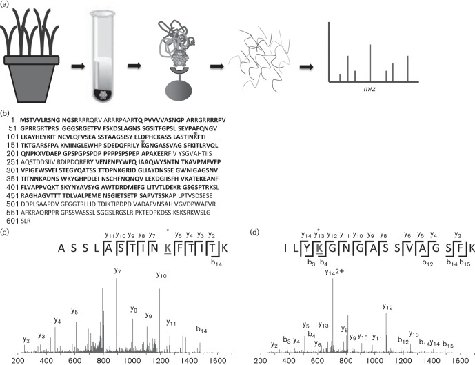
Fig. 1.
Discovery proteomics workflow identifies acetylated lysine residues in the CP of CYDV-RPV. (a) Oat tissue was homogenized and virions were enriched by density centrifugation followed by immunoprecipitation. The beads were subjected to tryptic digestion and the resulting peptides were analysed by tandem MS. (b) Tryptic coverage of the RTP (including the CP). Acetylated lysine residues are indicated with a triangle above the residue. Tryptic peptides are in bold type. (c) Tandem mass spectrum produced using collision induced dissociation (CID) in the Velos ion trap of the peptide ASSLASTINAcKFTITK. (d) Tandem mass spectrum of the peptide ILYAcKGNGASSVAGSFK, produced using CID in a Velos ion trap.

