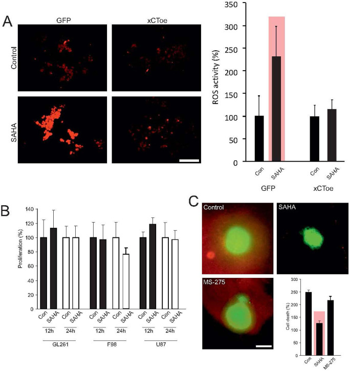
Figure 5. SAHA alleviates xCT-dependent cell death.
(A), Treatment of F98 glioma cells with SAHA is associated with tumor cell stress, as measured by ROS-accumulation. Quantification of ROS levels shows a significant increase in ROS after SAHA treatment. Stable over-expression of the xCT transporter leads to silencing of this effect and ROS activation through SAHA was no longer in evidence. These data show that SAHA induced suppression of the xCT transporter specifically lead to ROS activation, thereby resulting in cell stress. (B), However, the described cell stress was not accompanied by growth inhibition, which is analyzed by MTT assay. (C), F98 glioma cells were implanted on living organotypic brain slices and tumor-induced cell death analyzed through live imaging. Malignant gliomas induced neuronal cell death in surrounding healthy brain parenchyma, while treatment with the HDAC-inhibitor SAHA blocked neurodegeneration completely. Corresponding treatment with the HDAC inhibitor MS-275 does not lead to neuroprotection. Statistical analysis of neuronal cell death shows a significant reduction in PI positive cells after treatment with SAHA, whereas treatment with MS-275 did not achieve this effect. Statistical significance is calculated with the Student's t-test (mean ± s.d., *P < 0.05, n ≥ 6 per group).

