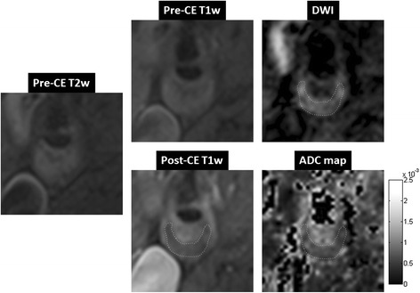
Figure 7.

A case study of a symptomatic subject with an atherosclerotic plaque of 70% stenosis in the right internal carotid artery. Pre-contrast T2-weighted image showed slightly hyper-intense signal throughout the plaque area. Pre-contrast T1-weighted image was iso-intense in the plaque area. Post-contrast T1-weighted image showed a clear hypo-intense area within the plaque, a typical LRNC appearance, surrounded by enhanced fibrous plaque tissue. DWI (b = 300 mm2/s) using DP-TSE showed a hyper-intense region, i.e. low diffusion, that spatially matched to the LRNC area in the post-contrast T1-weighted image. ADC map showed an area with low diffusion (0.62 ± 0.15 × 10⁻³ mm2/s) within the plaque that spatially matched to the LRNC finding in the post-contrast T1-weighted image.
