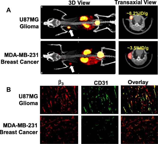
Figure 7.

(A) 3D and transverse views of SPECT/CT images of the athymic nude mice bearing U87MG glioma and MDA-MB-231 breast tumor xenografts. Each animal was administered with ∼37 MBq of 99mTc-P6G-RGD2. SPECT/CT study was designed to illustrate their potential utility for tumor imaging. (B) Selected microscopic images (Magnification: 200×) of the tumor slice stained with hamster anti-integrin β3 (red) and rat anti-CD31 (green) antibodies. Yellow or orange color (red integrin β3 staining merged with green CD31 staining) indicates the presence of integrin αvβ3 on the tumor neovasculature.
