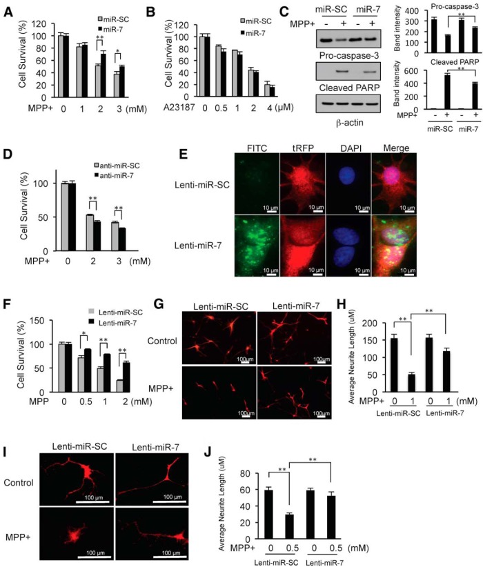
Figure 1.
MiR-7 protects against MPP+-induced cell death. A, SH-SY5Y cells were transfected with either pre-miR-SC or pre-miR-7 at a concentration of 50 nm. After 48 h, cells were challenged with MPP+ for 24 h, and cell survival was assessed. The survival of cells without MPP+ treatment was set at 100%. Ectopic expression of miR-7 without MPP+ exposure did not impact cell survival. B, SH-SY5Y cells were transfected with either pre-miR-SC or pre-miR-7, and were treated with A23187, and cell survival was measured. C, Cells were transfected as in A, and MPP+ (2 mm) was added for 24 h. Total cell lysates were subjected to Western blot analysis with antibodies against pro-caspase-3 or a cleaved form of PARP. Quantification of band intensities was calculated from three different experiments by normalizing to β-actin using NIH ImageJ software. D, Cells were transfected with either miR-7 inhibitor (anti-miR-7) or control (anti-miR-SC). After 24 h, MPP+ was added for another 24 h, and cell survival was measured. Experiments were performed in triplicate. E, FISH for miR-7 in differentiated ReNcell VM cells transduced with lenti-miR-7 or lenti-miR-SC. Cells transduced with lenti-miR-7 (tRFP) showed a high level of miR-7 expression (fluorescein isothiocyanate, FITC). F, Differentiated ReNcell VM cells infected with lenti-miR-7 or lenti-miR-SC were challenged with MPP+ at the indicated concentrations for 24 h, and cell survival was measured. For this experiment, infection efficiency was nearly 100%. G–J, Measurements of neurite length. Differentiated ReNcell VM cells (G, H) and mouse primary neurons (I, J) were transduced with lenti-miR-7 or lenti-miR-SC followed by MPP+ treatment for 24 h. For differentiated ReNcell VM cells, 20 tRFP-positive cells were used for measuring neurite length. For mouse primary neurons, 12 tRFP-positive neurons were used. Quantitative data are shown as the mean ± SEM. *p < 0.05; and **p < 0.01.

