
Figure 7.
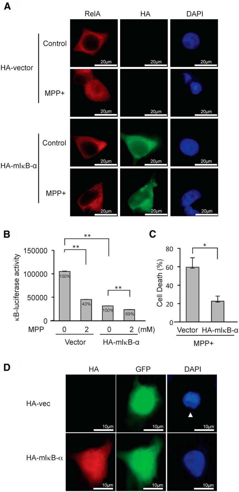
mIκB-α inhibits MPP+-induced NF-κB suppression and cell death. A, The expression of HA-mIκB-α inhibits nuclear localization of RelA upon MPP+ treatment. Endogenous RelA is stained as red fluorescence, and HA-mIκB-α is stained as green fluorescence. B, Expression of HA-mIκB-α inhibits MPP+-induced NF-κB suppression. Relative percentage values are calculated against non-MPP+ control. C, Expression of HA-mIκB-α inhibits MPP+-induced cell death. A 10 times higher amount of plasmid expressing HA-mIkB-α was cotransfected with EGFP-C1 into SH-SY5Y cells to ensure that nearly all GFP-positive cells express HA-mIκB-α. Cells having fragmented nuclei were counted in eight randomly selected fields comprising 10–30 cells among GFP-positive cells following treatment with 2 mm MPP+ for 24 h. The data represent the mean ± SE. *p < 0.05. D, A representative picture demonstrating an apoptotic nucleus (arrowhead) in a cell transfected with HA-vector control, whereas a nucleus from a cell expressing HA-mIκB-α appears intact following MPP+ exposure.