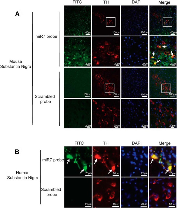
Figure 9.
Physiological expression of miR-7 in dopaminergic neurons in human and mouse brain. A, B, Substantia nigra sections from mouse (A) or human (B) brains were hybridized with miR-7 (green) or a scrambled probe along with immunostaining for TH (red). Nuclei were counterstained with DAPI (blue). Solid arrows indicate the expression of miR-7 in TH-positive neurons. Boxed areas are enlarged in the following panel.

