Figure 1.
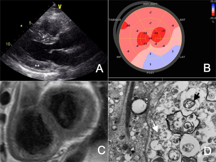
(A) Transthoracic echocardiography demonstrating a normal left ventricular cavity with increased septal (*) and posterior wall (**) thickness. (B) Bull's eye plot of the left ventricle obtained by two-dimensional speckle tracking echocardiography displaying reduced myocardial deformation in all segments (normal −18±2%). (C) Cardiac MRI after gadolinium injection without evidence of late contrast enhancement. (D) Lamellar bodies (black arrow) and curvilinear bodies (white arrow) viewed on transmission electron microscopy.

