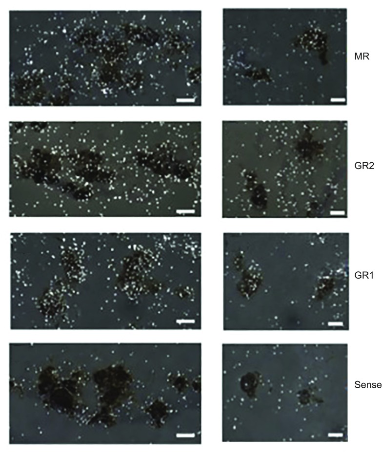
Fig. 2.
Cortisol receptor expression within gonadotropin releasing hormone (GnRH1) neurons. Photomicrographs of the preoptic area (POA) containing diaminobenzidine (DAB)-labeled GnRH1 neurons (brown). Left and right panels show separate locations in the field. The receptors responsive to cortisol are labeled by in situ hybridization using 3H-thymidine where silver grains show double label with mineralocorticoid receptor (MR) and glucocorticoid receptor GR1, but not GR2. In situ labeling with a sense probe for GR1 was used as a control. Scale bars, 20 μm.

