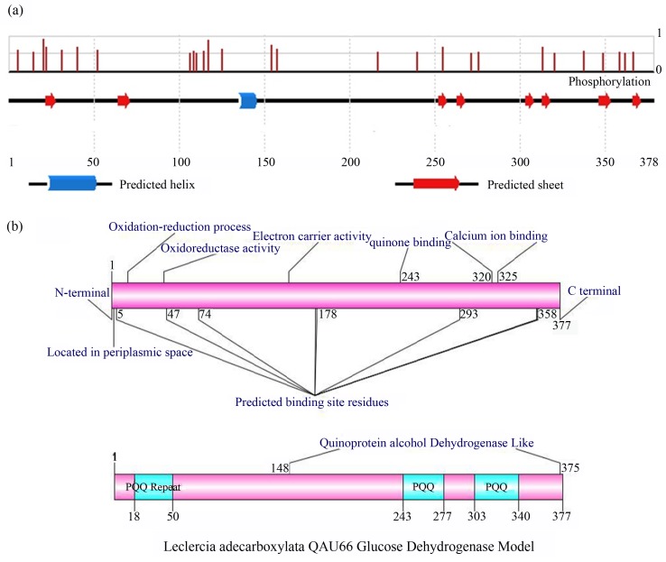
Figure 3.
(a) PSIPRED schematic presentation of secondary structure of QAU-66 GDH and position dependent features of Phosphorylation. The line height of the Phosphorylation features reflects the confidence of the residue prediction. The blue color represents the α-helix and red colour represents the β-strands. (b) Functional domains of QAU-66 GDH (A & B) predicted by INTERPROSCAN and COFACTOR respectively, and then drawn by DOG 2.0 (illustrator of protein domain structures).

