Figure 1.
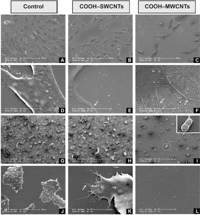
Scanning electron microscopy micrographs of hFOB (A–F) and mESC (G–L) cell lines after 3 days of culture on control and carboxyl-modified CNTs.
Notes: hFOB cells growing on control, COOH–SWCNT, and COOH–MWCNT substrates (A, B, and C, respectively); establishment of cytoplasmic nanoprotrusions on control, COOH–SWCNT, and COOH–MWCNT scaffolds, as seen at higher magnification (D, E, and F, respectively); and mESC colonies adhering to control scaffolds, COOH–SWCNTs, and COOH–MWCNTs (G, H, and I, respectively). Inset (I) shows abnormal morphology of colonies grown on COOH–MWCNTs magnified; higher magnifications depict establishment of nanoprotrusions on control and COOH–SWCNT substrates (J and K, respectively); inferred increase in nanoroughness introduced by COOH–MWCNTs (L). White arrows in Figures 1E, F and K depict the establishment of cytoplasmic prolongations of the cells to adhere to the scaffold.
Abbreviations: hFOB, human fetal osteoblast; mESC, murine embryonic stem cell; CNTs, carbon nanotubes; COOH–SWCNTs, carboxyl-modified single-walled CNTs; COOH–MWCNTs, carboxyl-modified multi-walled CNTs.

