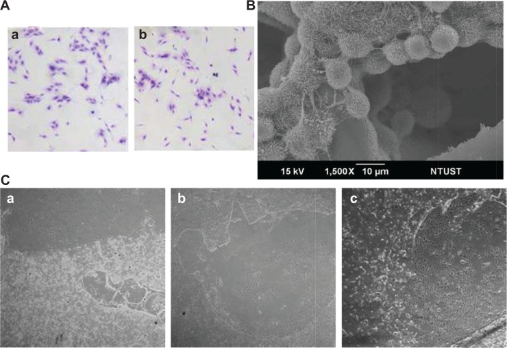
Figure 2.
Osteoblast adhesion and biocompatibility of chitosan nanofibers.
Notes: Mouse osteoblasts were seeded overnight in 6 cm tissue culture plates coated with chitosan nanofiber scaffolds. Cell adhesion was assayed using a crystal violet staining protocol (A): (a) CS film; (b) CS nanofibers. Also, adhesion of mouse osteoblasts onto chitosan nanofiber scaffolds was further analyzed using scanning electron microscopy (B). (C) After culturing mouse osteoblasts on chitosan nanofiber scaffolds for (a) 1, (b) 3, and (c) 5 days, the biocompatibility of the electrospun scaffolds was observed and photographed using light microscopy. 40× magnification.
Abbreviations: CS, chitosan; NTUST, National Taiwan University of Science and Technology.

