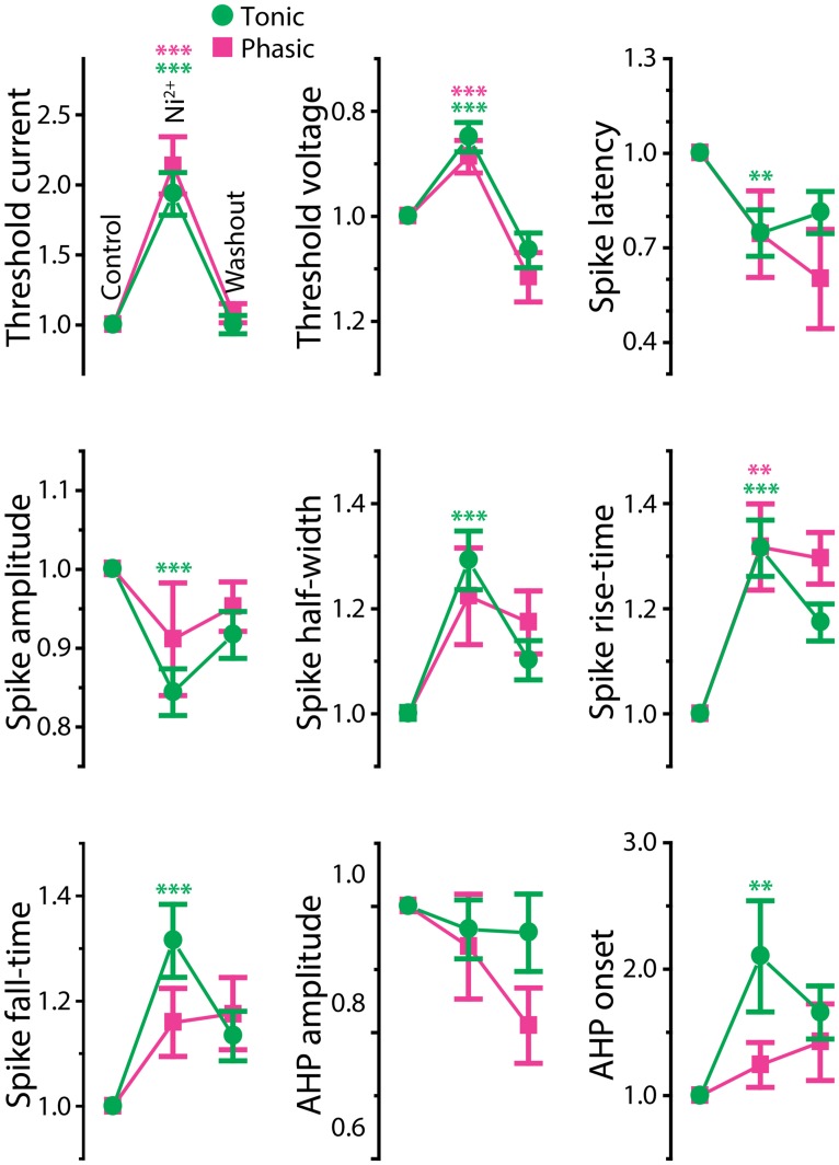
FIGURE 6.
Effects of Ni2+ on spike shape of tonic and phasic neurons. Spike parameters of tonic (green circle) and phasic (magenta square) neurons before (control; left), during (Ni2+; middle) and after (washout; right) Ni2+ application. Although measurements are reported as relative value to control, statistical significance was evaluated using raw values. Asterisks indicate significant difference between control and Ni2+ (green: tonic neurons, magenta: phasic neurons; double and triple asterisks represent p < 0.01 and p < 0.001, respectively, Holm–Sidak multiple comparison test following one-way repeated measures ANOVA). Ni2+ application significantly reduced spike amplitude and increased spike duration (half-width, rise-time, fall-time, and AHP onset) in tonic neurons. In phasic neurons, Ni2+ also increased spike rise-time; however, it did not significantly affect spike amplitude, fall-time, half-width, and AHP onset. Note that the treatment increased threshold current and depolarized threshold voltage similarly in tonic and phasic neurons.

