


Figure 2.
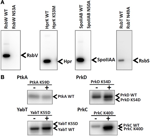
Inactivation of catalytic residues in all tested B. subtilis kinases. (A) Autoradiography images of the in vitro phosphorylation assays of RsbV, Hpr, SpoIIAA, and RsbS phosphorylated by their respective kinases: RsbW, HprK, SpoIIAB, and RsbT. Final protein concentrations in the assays were: 10 μM RsbV with 5 μM RsbW WT or RsbW N53A, 10 μM Hpr with 5 μM HprK/P WT or HprK K53M, 10 μM SpoIIAA with 1 μM SpoIIAB WT or SpoIIAB N50A, 3 μM RsbS with 3 μM RsbT WT or RsbT N49A. Bands corresponding to phosphorylated substrates are indicated by arrows. (B) Autoradiography images of intermolecular autophosphorylation assays (using incorporation of 32P) between wild type kinases PtkA, PrkC, PrkD, and YabT and their respective mutated versions PtkA K59D, PrkC K40D, PrkD K54D, and YabT K55D. Final protein concentrations in the in vitro phosphorylation assay were: 1 μM PtkA WT, 1 μM TkmA, 1 μM PtkA K59D, 1 μM PrkD WT, 1.5 μM YbdM K54D, 0.5 μM YabT WT, 0.5 μM YabT K55D, 0.5 μM PrkC WT, and 0.6 μM PrkC K40D. Wild type proteins were present on all lanes, and the presence or absence of mutant proteins is indicated with +/− above each lane. In order to be able to separate the WT kinases from the mutant versions, PtkA WT, PrkD WT, YabT K55D, and PrkC WT were fused with a 6xHis-tag and the respective mutant versions were Step-tagged fusions. Bands corresponding to phosphorylated kinases are indicated by arrows.