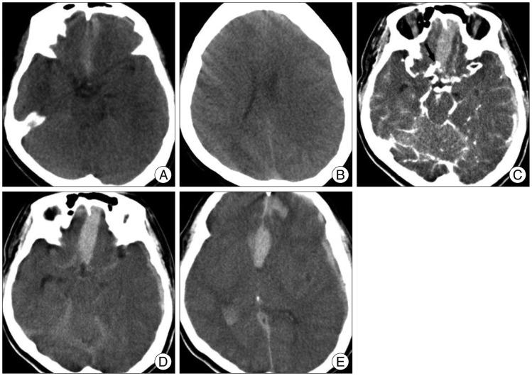
Fig. 1.
A : Initial plain CT scan shows thin SAH and subdural hematoma. B : There is no cavum septum pellucidum on the vertex cut. C : Source image of CTA reveals saccular aneurysms at the anterior communicating artery (arrow) and the left middle cerebral artery. D and E : Delayed images immediately after CTA show extensive SAH associated with intracerebral and intraventricular hemorrhages as well as blood within the septum pellucidum. CTA : computed tomography angiography, SAH : subarachnoid hemorrhage.

