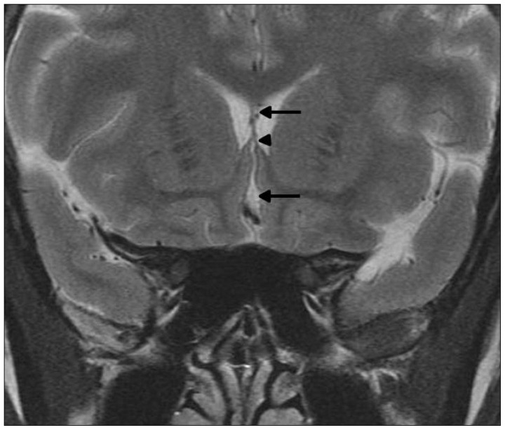
Fig. 4.
Coronal T2-weighted magnetic resonance image shows the thin strip of white matter of the lamina rostralis (arrowhead). This separates the anterior interhemispheric cistern (lower arrow) from the space between the leaves of the septum pellucidum (upper arrow) as well as the floor of the frontal horns in the mature brain.

