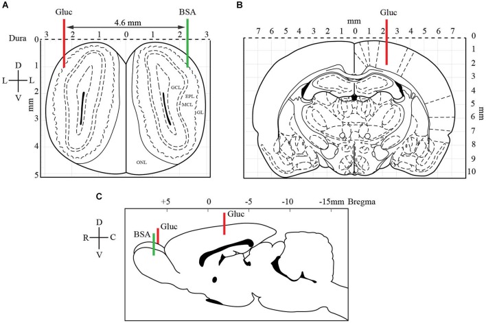
Figure 2.
Localization of glucose-oxidase biosensors (red) and control BSA sensor (green) electrodes in OB (A, C) and somatosensory cortex (B, C). At the OB level (A), two electrodes were implanted symmetrically (coordinates: AP +6.5 mm from Bregma, M/L −2.3 mm and D/V −1.0 mm from dura). A glucose-oxidase biosensor (Gluc, red line) was positioned in one bulb and a control BSA sensor (BSA, green line) in the other bulb. The latter was set measure nonspecific variations in oxidation current. In the somatosensory cortex (B), contralateral to the OB glucose sensor, the second glucose-oxidase biosensor was inserted (coordinates: AP −3.5 mm from Bregma, M/L +2.3 and D/V −2.3 mm from dura). The coronal drawings are from the Paxinos and Watson (2007) atlas, plates 1 and 27 (EPL: external plexiform layer, GCL: granular cell layer, GL: glomerular layer, MCL: mitral cell layer).

