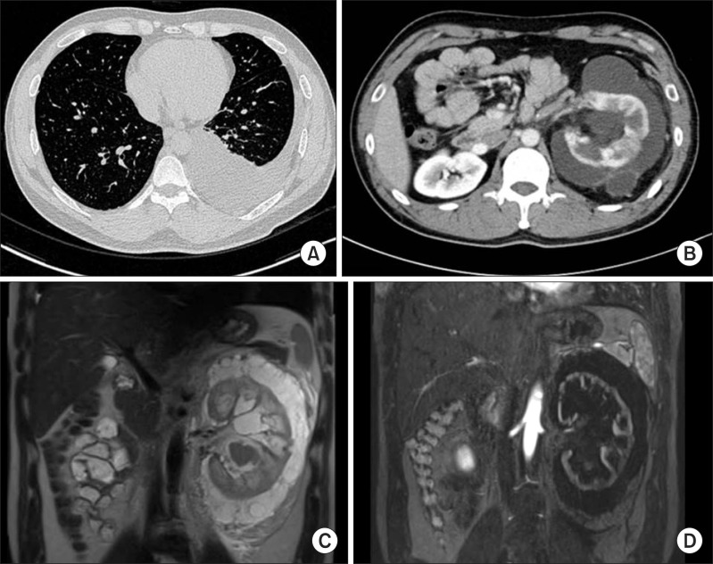
Fig. 1.
Radiologic images of perirenal lymphangiomatosis. (A) Chest computed tomography (CT) reveals pleural effusion of the left pleural cavity. (B) An enlarged left kidney has decreased radio-contrast enhancement with septated perirenal cystic fluid collections on abdominal CT. (C) High signal intensity is found around the left kidney with enlarged pelvo-calyceal system on T1-weighted magnetic resonance (MR). (D) Cortical thinning and irregularity of the left kidney is demonstrated in contrast enhanced T2-weighted MR.

