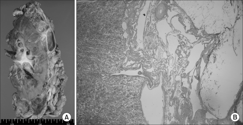
Fig. 2.
Gross and microscopic findings. (A) Multilocular cystic mass is enclosing the kidney. The mass consists of multiple cysts having thin fibrous walls and contains clear serous fluid. (B) Low power (×40) microscopic view shows multiloculated cystic spaces with various caliber of muscular and fibrous walls and without definite epithelial lining on H&E staining.

