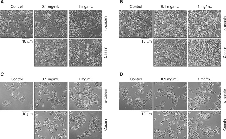
Fig. 3.
The effects of α-casein and casein on morphology. The morphological changes of various cells were observed by light microscopy after treatment with α-casein and casein under serum-free conditions for 72 hours. Under control conditions (vehicle: NaOH), PC-3 cells appeared to have a typical phenotype, featured with round nuclei and homogeneity. (A) After treatment with casein (or α-casein), PC-3 cells showed the following different morphological changes: 1) increase in cell volume, 2) more cohesion, and 3) marked increase in cell number. (B) Likewise, LNCaP cells showed increased cellular adhesion. (C) Treated MCF7 cells showed no morphological changes as compared to the untreated cells. (D) RWPE-1 cells also showed an increase in cellular adhesion without the changes in cell volume and number.

