Fig. 3.

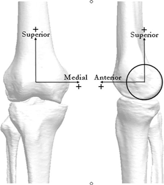
Representative bone geometry model derived from the CT scan depicting coordinate axes used to define the reference position of the tibia and femur: medial–lateral (ML), superior–inferior (SI), and anterior–posterior (AP)

Representative bone geometry model derived from the CT scan depicting coordinate axes used to define the reference position of the tibia and femur: medial–lateral (ML), superior–inferior (SI), and anterior–posterior (AP)