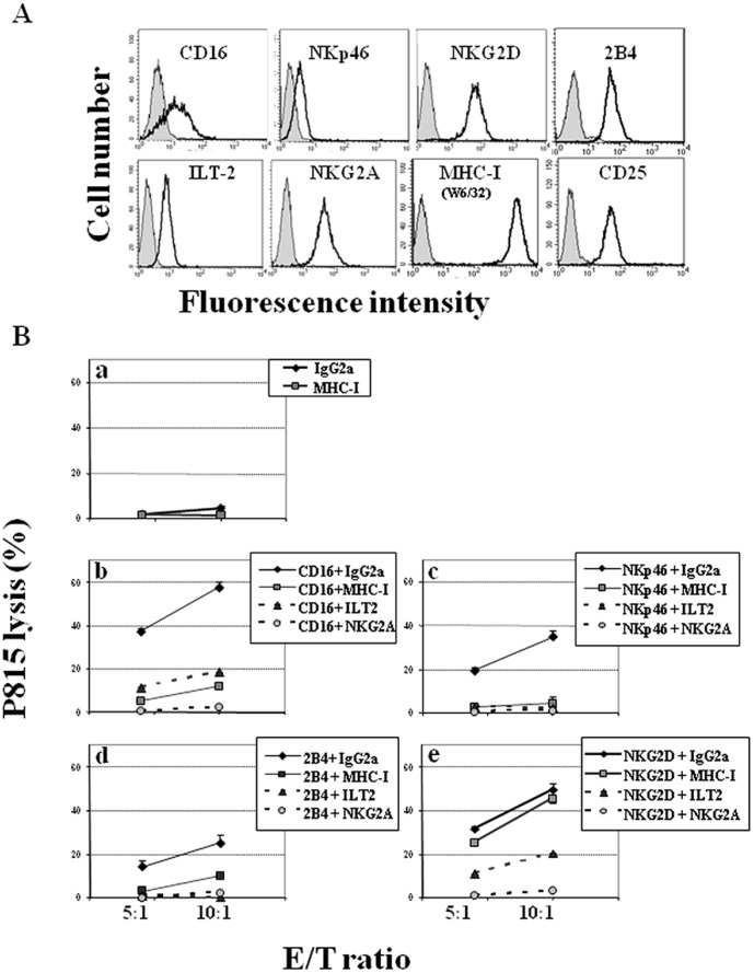
Figure 1. MHC-I engagement selectively inhibits cytotoxicity on NKL cells.
(A) Exponentially growing NKL cells (see Material and Methods section) were phenotyped by flow cytometry. Filled histograms represent isotype control and open histograms represent surface receptor stained cells. (B) NKL cells were co-cultured with 51Cr-P815 cells in the presence of mAb: IgG2a isotype control or anti-MHC-I (a), against KAR (CD16 (b), NKp46 (c), 2B4 (d), and NKG2D (e)), plus control IgG2a or anti-MHC-I, anti-ILT2 or anti-NKG2A inhibitory receptors. The figure depicts one representative assay out of three performed with similar results.

