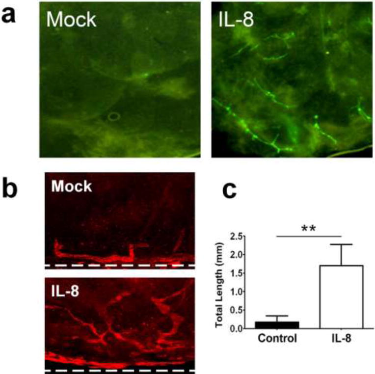
Figure 4. Promotion of in vivo lymphangiogenesis by IL-8.

(a) Matrigel plug assay showing induced in vivo lymphangiogenesis by IL-8. To directly visualize lymphatic ingrowth, lymphatic-specific GFP mice [33] were employed for matrigel plug assay. Growth factor-depleted matrigels premixed with Mock (PBS) or IL-8 (50 ng/ml) were subcutaneously inoculated into the left or right flank of the same mouse (two matrigel plugs per mouse, 5 mice per group). After two weeks, lymphatic vessel sprouting into the matrigels was directly visualized under a fluorescent stereoscope. Increase in the lymphatic vessel sprouting was quantified and shown in Supplemental Figure 3. This experiment was repeated twice and comparable results were obtained. (b) Mouse cornea micropocket assay was performed using wild-type mice. Lymphatic-specific LYVE-1 immunofluorescent staining demonstrates prominent ingrowth of new lymphatic vessels into the cornea in 14 days after IL-8 pellet implantation. Ten mice were used for each group (n=10). Dotted lines indicate demarcation between the cornea and conjunctiva. Original Magnification: ×50. (c) The outcome of the micropocket assay was quantitatively displayed as total lymphatic length ± standard error. **, p < 0.01.
