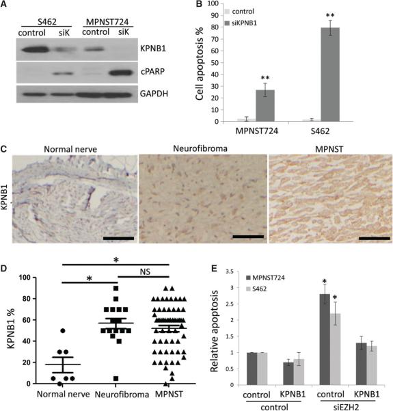
Figure 5.
KPNB1 knockdown induces cell apoptosis in MPNST cells and KPNB1 force expression rescued MPNST cell apoptosis induced from EZH2 knockdown. (A) Western blot analyses of KPNB1, cPARP and GAPDH in MPNST724 and S462 cells transfected with control or KPNB1 siRNA (siK). (B) Cell apoptosis assay of MPNST724 and S462 cells transfected with control siRNA or KPNB1 siRNA (siKPNB1). Data are shown as mean ± SD; n = 3; **p < 0.005, Student's t-test. (C) Representative immunohistochemical staining image of KPNB1 in normal nerve, neurofibroma and MPNST specimens on a TMA. Scale bars = 100 μm. (D) Scatter plots depicting the nuclear and cytoplasmic KPNB1 expression percentage in normal nerve (n = 8), neurofibroma (n = 19) and MPNST (n = 68) specimens, as determined by IHC (*p < 0.05; NS, not significant; Student's t-test). (E) Apoptosis analyses of MPNST724 and S462 cells transfected with negative control or siEZH2 combined with negative control or KPNB1 over-expression vector. Data are shown as mean + SD; n = 3; *p < 0.05, Student's t-test.

