
Figure 3. Effect of methyl-β-cyclodextrin (βCD) on the distribution of AC5/6.
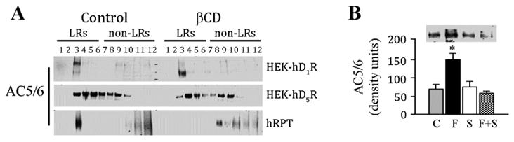
A. Cells from HEK-hD1R, HEK-hD5R, and hRPT were treated with vehicle (Control) or βCD (27%) for 1hr at 37°C. The cell lysates were then subjected to sucrose gradient centrifugation and the proteins (20µl/lane) from the sucrose gradient fractions (form 2 to 12 fractions) were immunoblotted with AC5/6 antibody. The distribution of AC5/6 in lipid rafts (LRs) and nonlipid rafts (non-LRs) is shown. n=3/group
B. The lipid raft marker caveolin 1 (Cav-1) co-immunoprecipitates with AC5/6 in hRPT cells
hRPT cells were grown to 90% confluence and then treated with vehicle (Control, C), fenoldopam (F, 1μmol/L), or SCH23390 (S) (5 μmol/L), or a combination of fenoldopam and SCH23390 (F+S). The cell lysates were immunoprecipitated with a polyclonal anti-Cav-1 antibody. Proteins from immunocomplexes were probed with a polyclonal anti-AC5/6 antibody. The immunoreactive bands were semi-quantified. Values are Mean ± SEM (n=4/group).
*P<0.05, vs. all others, one-way factorial ANOVA, Tukey post-hoc test. One immunoblot is shown.