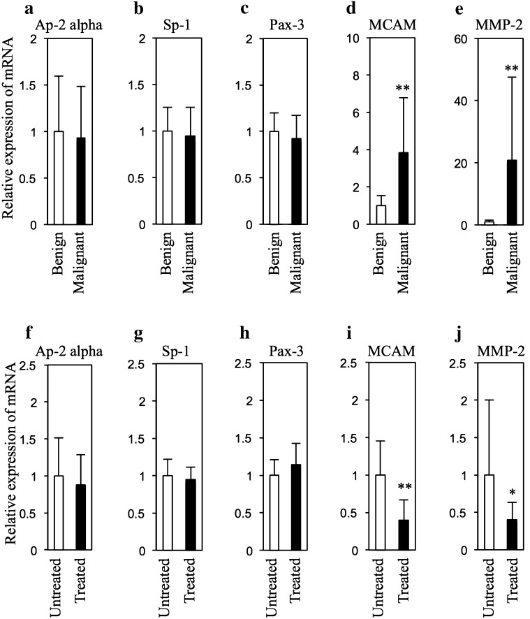
Fig. 1.
Expression levels Ap-2 alpha, Sp-1, Pax-3, MCAM and MMP-2 in tumors from HL-RET-mice. Relative mRNA expression levels (mean ± SD) of Ap-2 alpha (a, f), Sp-1 (b, g), Pax-3 (c, h), MCAM (d, i) and MMP-2 (e, j) in benign melanocytic tumors (n = 5) and melanomas (n = 5) (a–e) and in NEAPP-treated (n = 5) and untreated (n = 5) benign melanocytic tumors (f–j) in HL-RET-mice are presented. The mRNA expression levels were measured by quantitative PCR and adjusted by the mRNA expression level of hypoxanthine ribosyltransferase (Hprt) according to the method previously described [4]. The method of NEAPP irradiation for benign melanocytic tumors in HL-RET-mice for 11 weeks in this study was previously described [4]. Sequences of primers used in analysis of quantitative PCR are presented in Supplemental Table 1. Statistical significance was evaluated by Student’s t test (*p < 0.05, **p < 0.01). The experiments using recombinant DNA were approved by the Recombination DNA Advisory Committee of Chubu University (approval no. 12-04) and Nagoya University (approval no. 12-59, 13-59, 13-76). The experiments using animals were approved by the Animal Care and Use Committee of Chubu University (approval no. 2510052) and Nagoya University (approval no. 26317)

