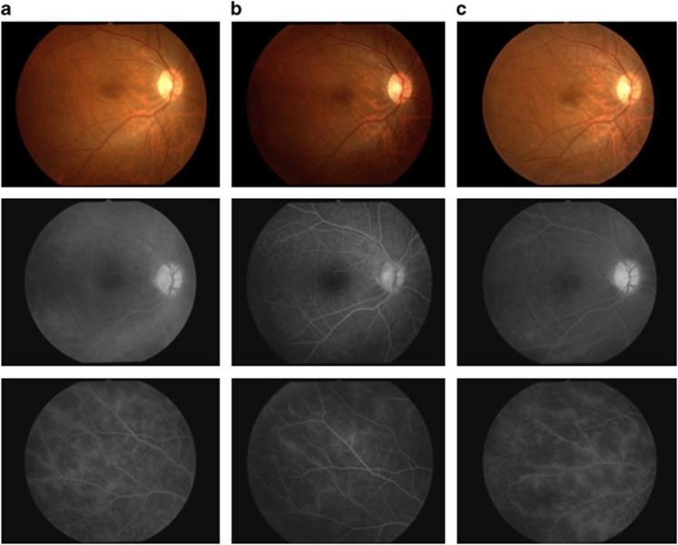
Figure 3.
Color fundus photographs and FA images of patient 5 at baseline and on infliximab therapy. Color fundus photographs and FA images are shown for the right eye of patient 5. All FA examinations were performed during times of clinical quiescence. (a) Fluorescein leakage from vessels at the optic disc, macula, and peripheral retina was observed before infliximab treatment (left upper and lower images). The vascular leakage score was 4.5 at this time (baseline). (b) Fluorescein leakage improved on infliximab therapy at the 6-month time point (middle upper and lower images). The vascular leakage score was 2.0. (c) However, FA leakage appeared to have increased at the end of 4 years (right upper and lower images) compared with at 6 months. The vascular leakage score was 4.0.

