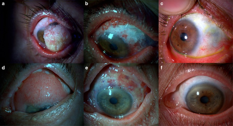
Figure 1.
(a) Preoperative picture of a 70-year-old female patient with squamous neoplasia involving two-thirds of the cornea. (b) Early postoperative picture showing the amniotic membrane. (c) Late postoperative (30 months) appearance of the eye. (d) Preoperative picture of an 84-year-old female with conjunctival intraepithelial neoplasia involving the cornea. (e) Early postoperative picture showing the ocular surface and amniotic membrane. (f) Late postoperative (24 months) appearance of the eye.

