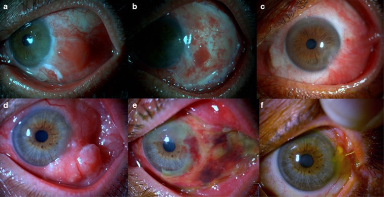
Figure 2.
(a) Preoperative picture of a 55-year-old male patient with squamous neoplasia. (b) Early postoperative picture showing the ocular surface. (c) Late postoperative (25 months) appearance of the eye to demonstrate the inferotemporal limbal stem cell deficiency. (d) Preoperative picture of a 60-year-old male with squamous neoplasia. (e) Early postoperative picture showing the amniotic membrane. (f) Late postoperative (18 months) appearance of the eye demonstrating mild superonasal symblepharon.

