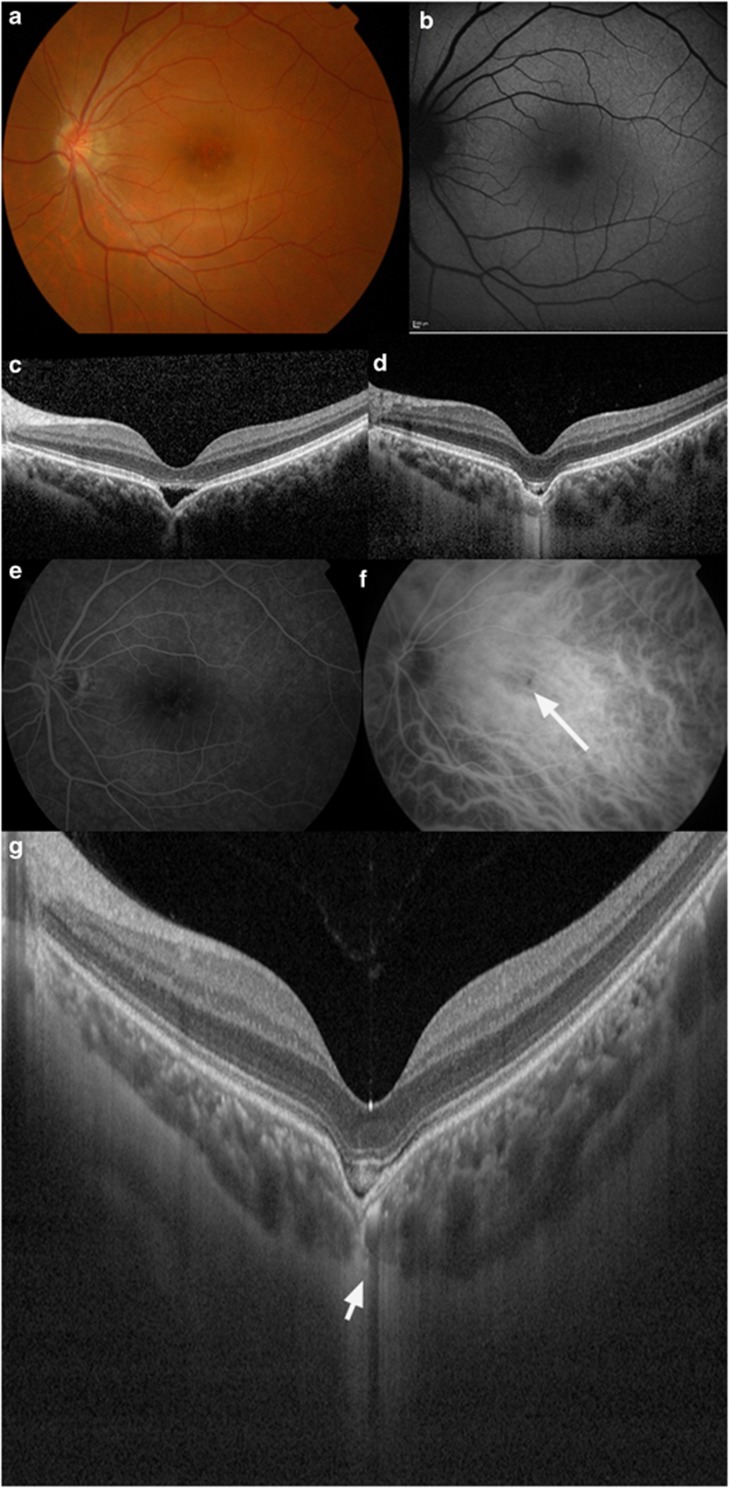
Figure 1.
A 20-year-old Chinese male with a recent history of CSC had a non-conforming FCE demonstrated on OCT that progressed to a conforming type over 6 months. (a) Fundal examination of the left eye revealed retinal pigment epithelial layer changes and mottling. (b) Fundal autofluorescence shows fine granular hyperautofluorescence in the macula. (c) Spectral-domain OCT (SD-OCT) revealed a non-conforming FCE in the macula of the left eye at presentation. (d) Repeat SD-OCT after 6 months follow-up revealed progression of the typical non-conforming choroidal excavation to the conforming type of choroidal excavation. (e) FFA did not show any abnormal findings. (f) ICG angiography revealed a hypofluorescent spot that corresponds to the FCE (see arrow). (g) SS-OCT showing a thinned-out choroid at the area of the excavation (see arrow) and an intact external limiting membrane and inner-segment/outer-segment junction. There was poor visualization of choroidal vasculature at the area of unusual thinned-out choroidal tissue and loss of contour of the outer choroidal boundary that appeared to be pulled inwards by this abnormal choroidal tissue. Note the presence of suprachoroidal space beneath this abnormal choroidal tissue. The choroidal–scleral interface was physiologically smooth, and the scleral was of normal curvature and thickness.

