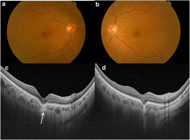
Figure 2.
An asymptomatic 56-year-old Chinese female with conforming FCE in both eyes. Color fundus photographs of the right eye (a) and left eye (b) show RPE mottling. (c) Swept-source optical coherence tomography (SS-OCT) of the right posterior pole showing thinned-out choroidal tissue just beneath the area of FCE (arrow). This thinned-out choroidal tissue had high internal reflectivity and there was poor visualization of both the medium and large-diameter choroidal vessels. There is loss of contour of the outer choroidal boundary, which appeared to be pulled inwards by this abnormal choroidal tissue. A suprachoroidal space was noted beneath this abnormal choroidal tissue and the choroidal–scleral interface was smooth. (d) SS-OCT of the left posterior pole showing similar findings to the right eye.

