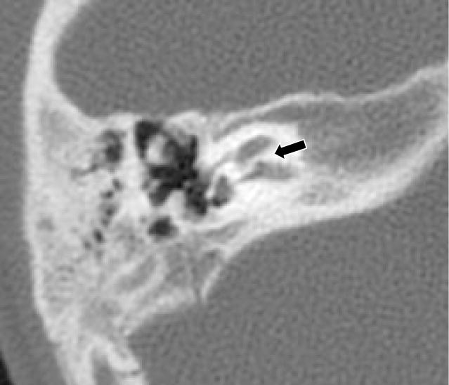
Figure 1.

Right Axial CT from patient 3 at the level of the cochlear canal shows lack of resorption of the bone that usually forms the cochlear canal (denoted by black arrow). Absence of the cochlear canal is usually indicative of aplasia of the cochlear nerve.
