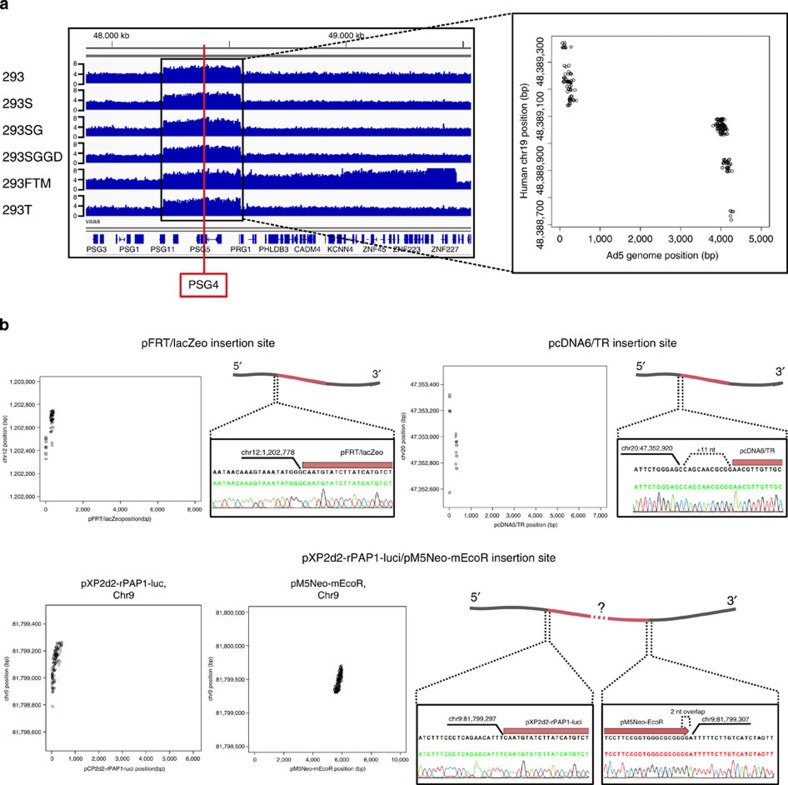
Figure 2. Plasmid insertion site detection.
(a) The Adenovirus 5 (Ad5) genome fragment is located in an 332.5-kb region on chr19 (48,221,000–48,553,500). This Ad5 sequence had been inserted and amplified in the 293 cell and the insertion and amplification have been maintained in the PSG4 gene of the whole 293 lineage. The Y-axis represents the genomic copy number. The dot plot in the right panel shows individual paired-reads aligning on the Ad5 genome (x axis) and chr19 (y axis). (b) Detection and confirmation of plasmid insertion sites in the 293FTM cell line. Four plasmids have been inserted into this cell line. Note the 11 additional bases inserted upstream of the pcDNA/TR plasmid (right panel), as well as the likely tandem insertion of pXP2d2-rPAP1-luci and pM5Neo-mEcoR plasmids on chr9 (bottom panel). Notably, we were unable to validate the plasmid–plasmid breakpoint of pXP2d2-rPAP1-luci and pM5Neo-mEcoR, probably due to the presence of stretches of homologous sequence in both plasmid sequences. Black sequence: consensus of several trace files, green or red sequences: derived from the representative trace file below the sequence. See also Supplementary File 4.

