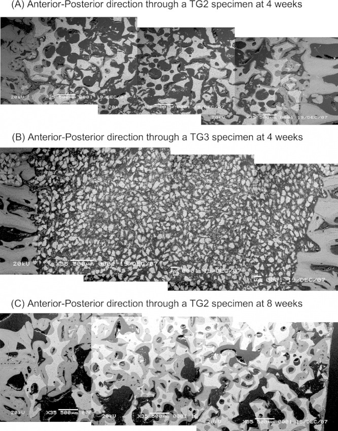
FIGURE 8.
A: A montage of SEM images through the defect of a TG2 specimen in the anterior/posterior direction at 4 weeks. B: A montage of SEM images through the defect of a TG3 specimen in the anterior/posterior direction at 4 weeks. In comparison to (A), the particle size is notably smaller. C: A montage of SEM images through the defect of a TG2 specimen in the anterior/posterior direction at 8 weeks.

