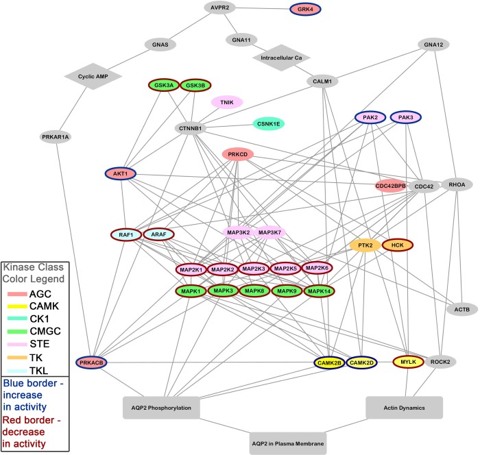
Fig. 4.
Protein-kinase network for the action of vasopressin in collecting duct cells was constructed from data in Table 1 using STRING (http://string-db.org/), followed by manual editing using Cytoscape (http://www.cytoscape.org/). Several additional proteins were added (see text) at the level of the STRING input to incorporate well-established knowledge about vasopressin signaling, viz., Avpr2, Aqp2, Prkar1a, Prkacb, RhoA, CDC42, Rock2, Gnas, Gna11, Gna12, Ctnnb1, Gsk3β, and Actb. Node colors indicate protein kinase family (see figure key). Node borders designate whether the protein kinase was inferred to increase (blue) or decrease (red) in activity. AQP2, aquaporin-2.

