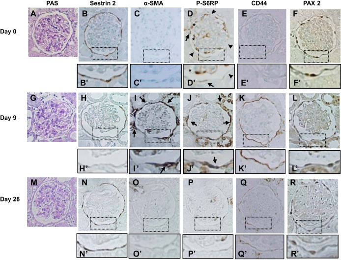
Fig. 6.
Immunohistochemical staining for each protein in PECs in a rat model of PAN nephropathy. Rats were injected with PAN, and kidney sections were examined on day 0 (A–F), day 9 (G–L), and day 28 (M–R). A, G, and M: PAS staining. B, H, and N: sestrin 2. Sestrin 2 expression decreased on day 9 (H and H′) but was restored on day 28 (N and N′). C, I, and O: α-SMA. Strong expression of α-SMA was detected around the basement membrane of Bowman's capsule on day 9 (arrows in I and I′) but disappeared on day 28 (O and O′). D, J, and P: P-S6RP. P-S6RP was detected weakly in some cells along Bowman's capsule (arrows in D and D′), but most cells were negative for P-S6RP expression on day 0 (arrowheads in D and D′). Strong expression of P-S6RP was observed in cells along Bowman's capsule on day 9 (arrows in J and J′), but the expression almost disappeared on day 28. Within the glomerular tufts, some cells showed strong staining for P-S6RP (* in D and D′). E, K, and Q: CD44. CD44 expression was increased on day 9 (K and K′) but returned to basal levels on day 28 (Q and Q′). F, L, and R: PAX2. PAX2 was detected in cells along Bowman's capsule in a nuclear localization. Original magnification: ×400.

