Fig. 1.
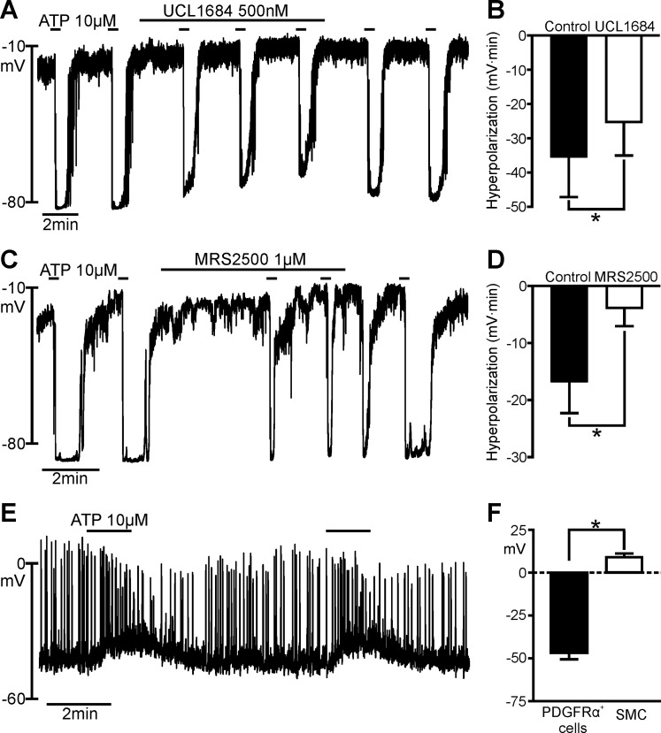
Effects of ATP on platelet-derived growth factor receptor-α-positive (PDGFRα+) cells and smooth muscle cells (SMCs). A and C: current-clamp recordings from PDGFRα+ cells in the whole cell configuration [current (I) = 0]. ATP (10 μM) elicited fast transient hyperpolarization with each application. Peak of hyperpolarization was about −80 mV [i.e., equilibrium potential for K+ (EK) under conditions of our experiments]. In A and C, hyperpolarization responses are reduced by UCL 1684 and MRS 2500; B and D show significantly reduced hyperpolarization responses. ATP responses recovered after washout of the inhibitors (A and C). B: summary of inhibitory effects of UCL 1684 on responses to ATP (n = 5). *P = 0.0260 (by paired t-test). D: summary of inhibitory effects of MRS 2500 on responses to ATP (n = 6). *P = 0.0073 (by paired t-test). Hyperpolarization responses in B and D are tabulated as area under response curves (mV·min). E: current-clamp recording from a SMC (I = 0) with perforated-patch, whole cell configuration. ATP (10 μM) elicited slowly developing depolarization in the SMC. F: summary of effects of ATP on PDGFRα+ cells and SMCs. ATP evoked opposite effects on resting membrane potentials (RMPs) of PDGFRα+ cells and SMCs. Average changes in membrane potentials were −46.7 ± 3.80 mV in PDGFRα+ cells (n = 20) and +13.5 ± 2.90 mV in SMCs (n = 7). *P < 0.0001 (by unpaired t-test).

