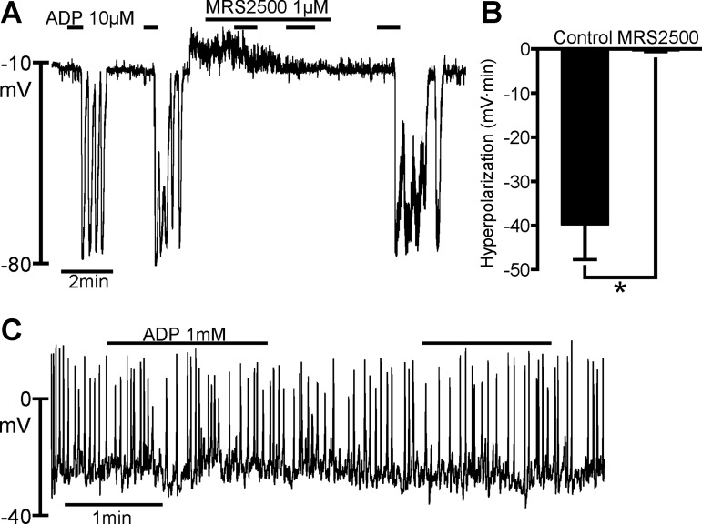
Fig. 2.
Effects of ADP on PDGFRα+ cells and SMCs. A: current-clamp recording from a PDGFRα+ cell in the whole cell configuration (I = 0). ADP (10 μM) elicited transient hyperpolarizations with repetitive applications; peak hyperpolarization reached about −80 mV. Hyperpolarization responses in this cell were oscillatory in nature. Hyperpolarization response was blocked by MRS 2500 (1 μM). ADP effects recovered within a few minutes after removal of MRS 2500. B: summary of inhibitory effects of MRS 2500 on ADP responses (n = 6). *P = 0.0041 (by paired t-test). Hyperpolarization responses in B are tabulated as area under response curves (mV·min). C: current-clamp recording from a SMC with perforated-patch, whole cell recording (I = 0). ADP (1 mM) had no effect on RMP of SMCs.

