Fig. 9.
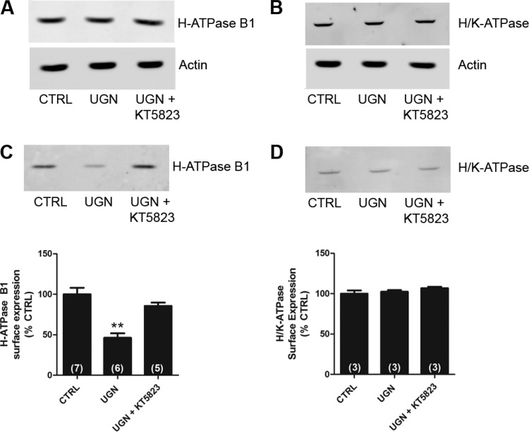
UGN decreases the surface expression of H+-ATPase B1 subunit in MDCK-C11 cells by a PKG-dependent mechanism. A and B: equivalent quantities (50 μg) of total cellular lysates from MDCK-C11 cells treated for 15 min with vehicle (CTRL), UGN, or UGN combined with the PKG inhibitor were subjected to SDS-PAGE and immunoblotting. A: representative immunoblotting using the monoclonal antibody against the B1 subunit of H+-ATPase. Actin was used as an internal control. B: representative immunoblotting using the monoclonal antibody against the β subunit of H+/K+-ATPase. Actin was used as an internal control. C and D: cell surface biotinylated proteins, after treatment with UGN or UGN + KT5823 (PKG inhibitor) for 15 min, were subjected to SDS-PAGE and immunoblotting. Immunoblot analyses were performed using a monoclonal antibody against the B1 subunit of H+-ATPase (C) or a monoclonal antibody against the β subunit of H+/K+-ATPase (D). The amount of H+-ATPase B1 subunit and H+/K+-ATPase β subunit were quantitated by densitometry. Number of experiments are indicated within the parenthesis. **P < 0.01 vs. CTRL.

