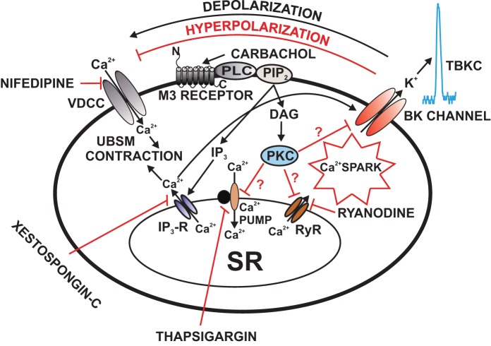
Fig. 4.
Illustration of the cellular mechanisms by which M3 receptors regulate BK channel function in UBSM cells. Activation of M3 receptors leads to IP3 and DAG production, via a pathway involving phospholipase-C (PLC) and PIP2. IP3 activates IP3 receptors, which releases Ca2+ from the SR. This IP3-induced Ca2+ release transiently activates the BK channels. Over time, depletion of SR Ca2+ upon activation of M3 receptors reduces Ca2+ spark activity, which leads to inhibition of TBKCs and depolarization of UBSM cell resting membrane potential causing activation of VDCC and thus increases UBSM contractility. DAG activates PKC, which may lead to direct inhibition of the SR Ca2+ pump and RyRs resulting in suppression of Ca2+ sparks and TBKCs. Pharmacological tools used in this study to inhibit cellular sources of Ca2+ are indicated. BK, large-conductance voltage- and Ca2+-activated K+ channel; DAG, 1,2-diacylglycerol; IP3, inositol triphosphate; M3 receptors, muscarinic receptors type 3; PIP2, phosphatidylinositol 4,5-bisphosphate; PKC, protein kinase-C; PLC, phospholipase-C; Ryr, ryanodine receptor; TBKCs, transient BK currents; SR, sarcoplasmic reticulum; UBSM, urinary bladder smooth muscle; VDCC, L-type voltage-dependent Ca2+ channel. This figure is based on information from (62, 112, 114).

