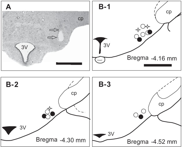
Fig. 5.
Histological illustrations showing the location of the microdialysis probe and the sensor in the posterior hypothalamus. A: a photomicrograph of the coronal section of the posterior hypothalamus showing the location of the microdialysis probe (indicated by arrows). B: location of microdialysis probe and glutamate sensor in three stereotaxic planes of rat brain through the posterior hypothalamus tuberomammillary nucleus (B1–3). The location of the tip of the microdialysis probe in animals for histamine (○) and glutamate-GABA (●) experiments; and glutamate sensor (stars). Scale bar = 1 mm. Scale bar in B-1 is same for B-2 and B-3. 3V, third ventricle mammillary recess; cp, cerebral peduncle.

