Fig. 1.
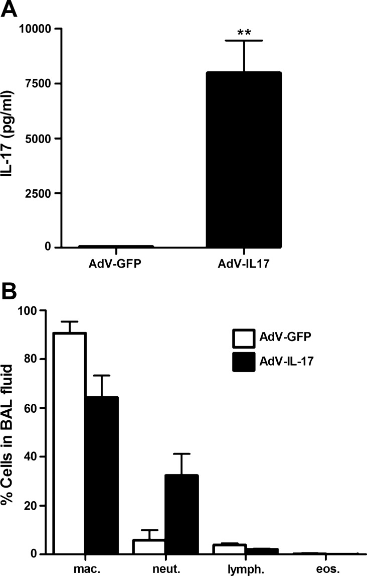
Expression of IL-17 and lung neutrophilia in IL-17-expressing recombinant adenovirus (AdV-IL-17)-treated mice. A: AdV-IL-17 or an equivalent amount (1×10⁸ pfu) of green fluorescent protein-expressing recombinant adenovirus (AdV-GFP) was delivered to C57BL/6 mice by oropharyngeal aspiration. One week posttreatment, bronchoalveolar lavage (BAL) was performed. The levels of IL-17 in the BAL fluid were determined by ELISA. Graph shows mean levels ± SE IL-17 in the first 0.8 ml aliquot of BAL fluid (**P < 0.01 vs. AdV-GFP group, n = 5 per group). B: differential cell counts of BAL cells. Cytospin samples of the cells recovered by BAL were stained with Hema 3 (Fisher). Cells were visualized by microscopy, and 200 cells were counted from each sample. Graph shows percentage of the indicated cell type (means ± SE) in the cells recovered from the BAL fluid from mice treated with AdV-IL-17 (solid bars) and AdV-GFP (open bars). P = 0.07, percentage neutrophils AdV-GFP vs. AdV-IL-17, n = 5 per group.

