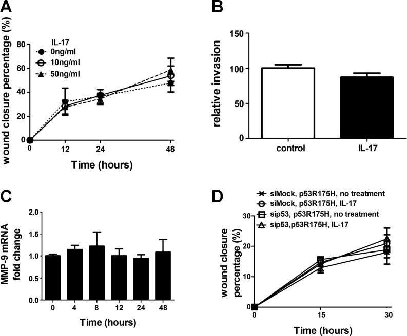
Fig. 6.
Mutant p53 alters IL-17-mediated promotion of migration and induction of MMP-9. A: effect of IL-17 on migration of mutant p53-expressing cells. Same as Fig. 4A except confluent mK-Ras-R172H-LE cells were assessed for migration in serum-free medium (●, dotted line) or serum-free medium supplemented with 10 (○, solid line) or 50 (△, dashed line) ng/ml IL-17. Graph shows percentage of wound closure vs. time. Data shown are means ± SE (n = 4). B: IL-17 does not promote invasion of mutant p53-expressing cells. Transwell migration assays (see Fig. 4B) were performed with mK-Ras-R172H-LE cells in serum-free media with (solid bar) or without (open bar) 10 ng/ml IL-17. Data shown are means ± SE (n = 4). C: total RNA was prepared from mK-Ras-R172H-LE cells at increasing times after treatment with 10 ng/ml IL-17. Graph shows the mean level of MMP-9 mRNA relative to β-actin (2−ΔΔCT method) at the indicated time after addition of IL-17 to the serum-starved cells. Data shown are means ± SE (n ≥ 5). D: same as Fig. 5C, except the cotransfected plasmid (pCMV-p53R175H) expressed the R175H mutant of human p53 in the K-Ras-LE cells with p53 knocked down. Graph shows percentage wound closure at the indicated times with mutant human p53 expression in cells transfected with mock siRNA (×) or p53 siRNA (□) in the absence IL-17 and in cells transfected with mock siRNA (○) or p53 siRNA (△) in the presence of 10 ng/ml IL-17. Data shown are means ± SE (n = 4).

