Figure 1.

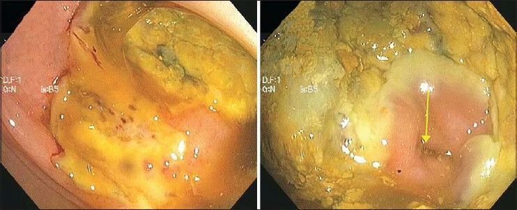
Colonoscopy images showing diffuse edema, severe ulceration and mucosal necrosis in the large intestine consistent with ischemic colitis. The region of the ileocecal valve is spared (arrow)

Colonoscopy images showing diffuse edema, severe ulceration and mucosal necrosis in the large intestine consistent with ischemic colitis. The region of the ileocecal valve is spared (arrow)