Fig 1.
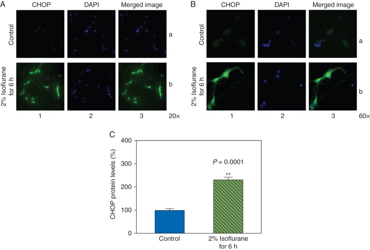
Isoflurane increases CHOP levels in the primary neurones. (a) Immunohistochemistry staining of CHOP (magnification 20 ×). (b) Immunohistochemistry staining of CHOP (magnification 60 ×). Column 1 is the image of CHOP (green), column 2 is the image of nuclei (blue), and column 3 is the merged image. Row a is the cells following the control condition and row b is the cells treated with 2% isoflurane for 6 h. (c) Quantification of the immunohistochemistry staining shows that the isoflurane treatment (green striped bar) increases CHOP levels compared with the control condition (blue bar).

