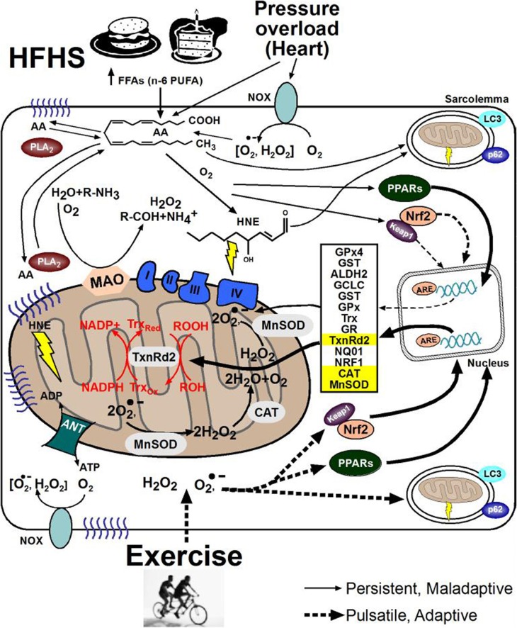
Figure 2.
Cell signaling pathways underlying ROS adaptation in striated muscle. The beneficial and deleterious consequences of oxidative stress coming from cardio-metabolic diseases (top) or exercise (bottom) in striated muscle at the subcellular level are shown here. Note the difference in designation of solid-line arrows for the persistent ROS coming from disease, compared to dashed-line arrows representing the pulsatile ROS coming from exercise. Formation of lipid peroxides and their derivative aldehydes such as 4-hydroxynonenal (HNE), along with their subsequent reactivity to cause protein carbonylation and carbonyl stress is depicted by yellow lightning bolts in cytosol and mitochondria. (NOX, NADPH oxidase; HFHS, high fat, high sucrose; PUFA, polyunsaturated fatty acids; FFA, free fatty acids; AA, arachidonic acid; PLA2, phospholipase A2; PPAR, peroxisome proliferator-activated receptor; ANT, adenine nucleotide translocase; COX IV, cytochrome oxidase IV; ATP, adenosine tri-phosphate; Nrf2, NF E2-related factor 2; NRF1, nuclear respiratory factor 1; GST, glutathione S-transferase; ALDH2, aldehyde dehydrogenase 2; GCLC, γ-Glutamylcysteine ligase catalytic subunit; GPx, glutathione peroxidase; NQO-1, NADPH-quinone oxido-reductase-1; GR, glutathione reductase; GSHt, total glutathione; Trx, thioredoxin; TxnRd2, thioredoxin reductase-2; MAO, monoamine oxidase; MnSOD, manganese superoxide dismutase; CAT, catalase).

