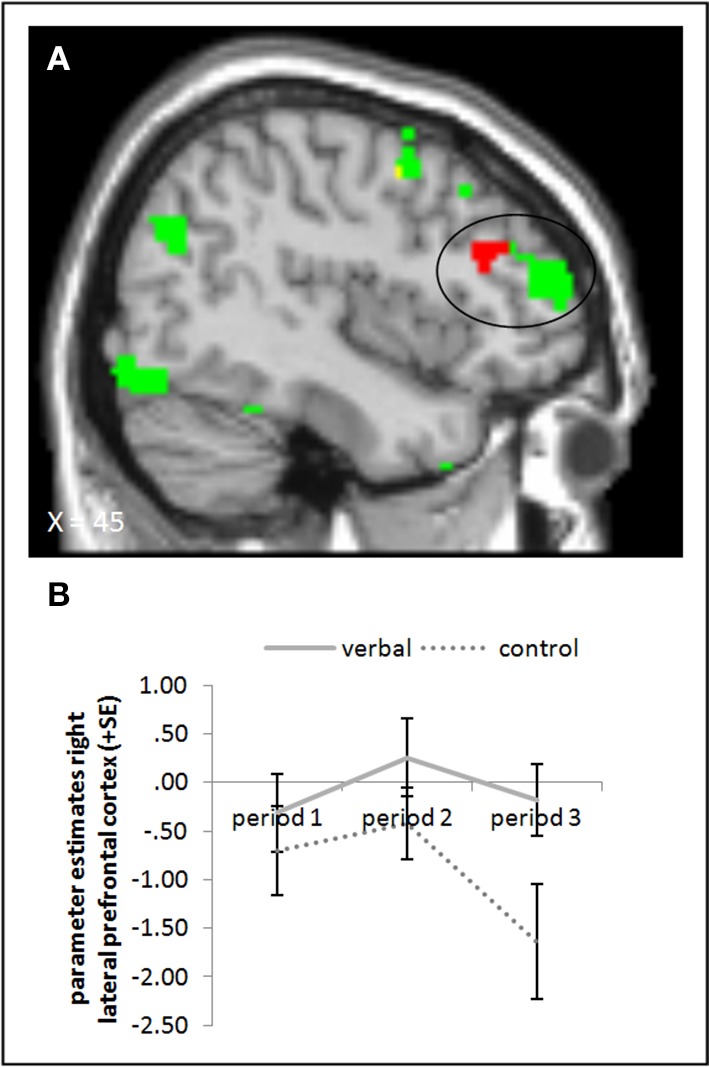
Figure 6.

(A) Activation for picture presentation was significantly higher in the right lateral prefrontal cortex in the verbal reward group compared to the control group in period 3 (whole brain uncorrected, p < 0.001, only clusters with a minimum of 10 activated voxels are shown). Activation clusters without baseline correction are shown in green, clusters with baseline correction are shown in red, activation overlaps are shown in yellow. (B) Parameter estimates are displayed for illustration.
