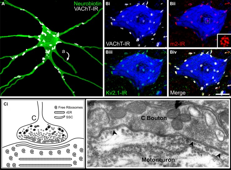
FIGURE 3.
The C-bouton synapse on mammalian α-motoneurons. (A) C-bouton synapses on intracellularly labeled and reconstructed adult rat lumbar α-MN are revealed by VAChT-IR (white). Large C-boutons densely innervate the soma and proximal dendrites of α-MNs but are absent from more distal locations. Also note that C-boutons are not located on motoneuron axons (indicated by “a”). (B) C-boutons, indicated by VAChT-IR (Bi,iv, white), are presynaptic to the muscarinic m2 receptor (Bii,iv, red) and large Kv2.1 clusters (Biii,iv, green). Note that m2 receptor immunoreactivity on the α-MN soma and proximal dendrites localize exclusively to C-bouton postsynaptic sites. (Bii) Inset shows subsynaptic fenestrated distribution of m2-IR. Images are confocal stacks of 12 × 1 μm Z-stacks with nissl stain (blue) to label adult rat neuronal somata. Scale bar is 20 μm. (C) Diagrammatic representation and electron micrograph of C-bouton ultrastructure in an adult rat. (Ci) Diagram illustrates densely packed, clear spherical or pleomorphic vesicles and abundant mitochondria. Closely apposed to the postsynaptic membrane is a 10–15 nm wide subsurface cistern (SSC) that is continuous with several lamellae of underlying rough endoplasmic reticulum (rER). Free ribosomal rosettes are typically visible in the subsynaptic region. (Cii) Electron micrograph of C-bouton synapse on an α-MN soma. Arrowheads indicate a SSC extending the entire appositional length of the bouton. Note key features present in electron micrograph illustrated in diagram (Ci).

