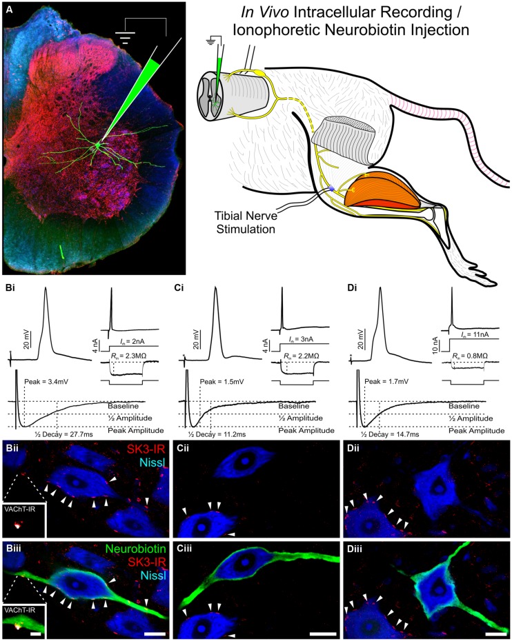
FIGURE 5.
Subset of rat lumbar α-motoneurons with SK3-IR have significantly longer AHP 1/2 decay time and increased amplitude. Data shown is review of previous study reported by Deardorff et al. (2013). (A) Diagrammatic representation of experimental paradigms. In an adult in vivo rat preparation, tibial α-MNs, identified by antidromic activation of the tibial nerve, were penetrated with a sharp recording electrode. Neuronal electrical properties were recorded and neurons were filled with neurobiotin (green) for post hoc identification. Spinal cord tissue was harvested and processed for SK3-IR. (B–D) Neuronal electrical properties are of α-MNs depicted in micrographs below. Asterisk (*) denotes stimulus artifact. Micrographs are single optical confocal sections through the soma of intracellularly labeled α-MNs (green) processed for SK3-IR (red) and the general neuronal stain nissl (Blue). Scale bars are 20 μm. (B) SK3-IR (+) (Bii and Biii arrowheads) α-MNs have long duration and large amplitude AHP, low rheobase, and high input resistance. Micrograph insets show VAChT-IR (White) C-bouton in apposition to an SK3-IR (+) cluster. Inset scale bar is 5 μm. (C,D) SK3-IR (-) α-MNs have short duration and small amplitude AHPs. However, even among these SK3-IR (-) cells, rheobase and input resistance show high variance along the continuum of α-MN properties. Please note the nearby SK3-IR (+) cells (C,Dii,iii arrowheads).

