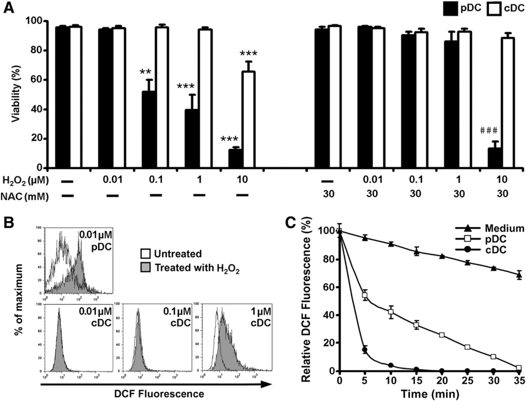
Fig. 1.
Sensitivity of pDCs and conventional DCs to oxidative stress. (A) Effect of exposure to H2O2 on the viability of pDCs and conventional DCs. Cells were treated with increasing concentrations of H2O2 for 24 h. In control experiments, cells were pretreated with an antioxidant, N-acetylcysteine, for 1 h and then cotreated with H2O2. After treatments, cells were stained with 7-AAD and the proportion of 7-AAD-negative (living) cells was assessed by flow cytometry. Percentages of viable cells are displayed. Data are presented as means ± SE of three individual experiments. **P<0.01, ***P<0.001 vs untreated control cells, ###P<0.001 vs cells pretreated with N-acetylcysteine. (B) Exposure to low-dose H2O2 (0.01 µM) increases intracellular ROS levels in pDCs, but not in conventional DCs. Cells were loaded with redox-sensitive H2DCFDA and treated with H2O2 for 2 h. Changes in intracellular DCF fluorescence were determined by flow cytometry. Results are representative of three independent experiments. (C) Consumption of H2O2 in cell culture medium of pDCs and conventional DCs. Cells were treated with 0.01 µM H2O2 and samples were withdrawn from the supernatant every 5 min to assay H2O2 content. To measure H2O2 levels, cell-free supernatant samples were mixed with H2DCFDA and fluorescence intensity was assessed by fluorimetry. Data are presented as means ± SE of three individual experiments. NAC, N-acetylcysteine; cDC, conventional DCs.

